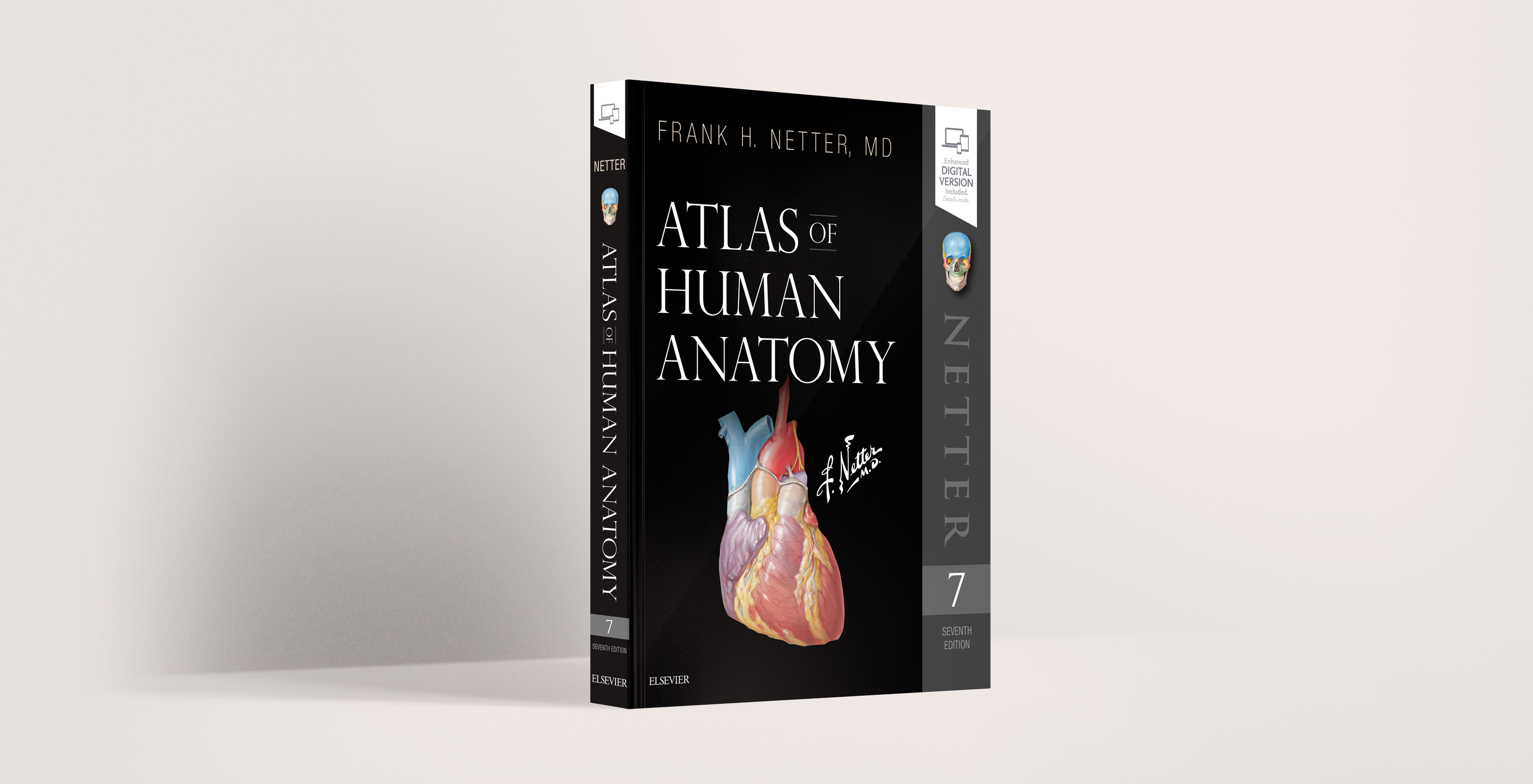
Netter’s Atlas of Human Anatomy is a masterful work of art and academia that allows you to learn by seeing. Filled with images originally illustrated by painter and renowned physician Dr. Frank Netter, this textbook is a vivid rendering of the human anatomy which is not only detailed and accurate, but also beautiful. Considered by students and professionals alike as a necessary resource and lifelong medical reference, this text is a must have.

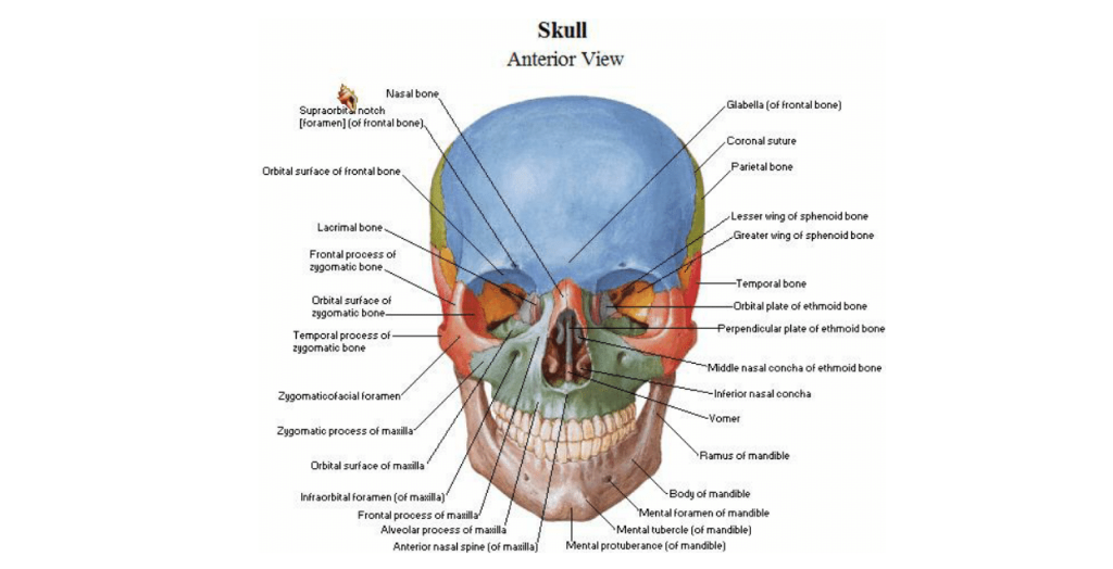
Netter’s Atlas gives a comprehensive overview of each system with images that elegantly depict every single anatomical structure. These images are intricately illustrated and generously labeled to help you develop a robust vocabulary and a strong understanding of the human body. While each structure is labeled to meet international terminology standards, commonly used clinical terms (e.g. Fallopian tubes) are included so that you’ll start to recognize them early on in your practice.

The latest (7th) edition of Netter’s Atlas comes with a few bonuses. The Overview section gives you a summary of all the key organ systems from head to toe, a summary of muscles, and case studies with x-rays, MRIs and CT scans. Each section includes a handy Clinical Reference table, which lists all the commonly injured anatomical structures so that you’ll start to recognize them during rotations.
The newest version of Netter’s Atlas also comes with a free interactive eBook that you can access online or offline. The eBook is powered with 3D models, videos, and enhanced images that come with a Test-Yourself assessment. This assessment lets you quiz yourself on any structure, region or system of your choosing. Test-Yourself gives you more than 300 questions to choose from as you make your way through the complexities of the human body.

Diese Website verwendet Cookies. Um sie abzulehnen oder mehr zu erfahren, besuchen Sie unsere cookie notice.