by Santiago Martínez-Jiménez, MD; Carlos S. Restrepo, MD
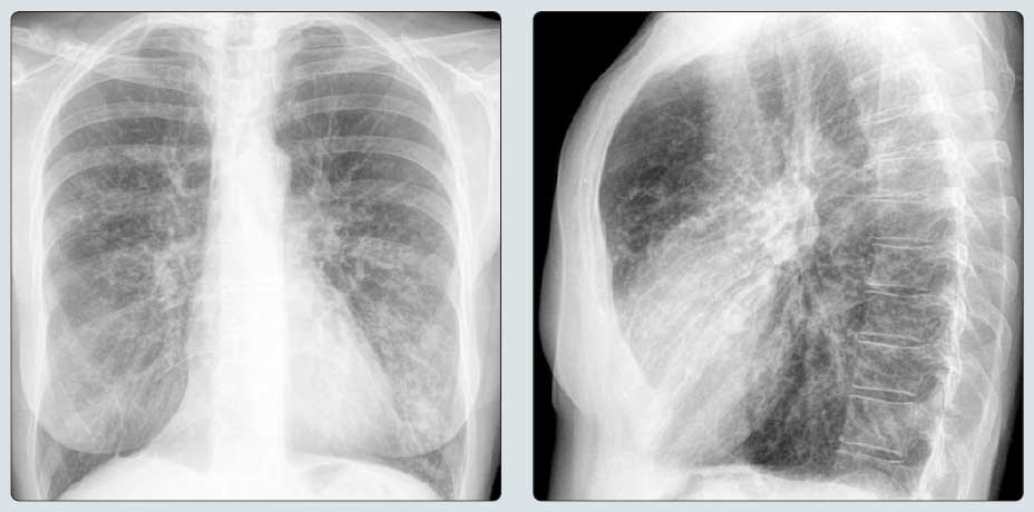
(Left) PA chest radiograph of a patient with longstanding asthma shows bilateral lung hyperinflation and nonspecific bilateral reticular opacities. (Right) Lateral chest radiograph of the same patient shows flattening of the diaphragm and enlargement of the retrosternal clear space, consistent with marked hyperinflation. Note diffuse peribronchial cuffing. While nonspecific, hyperinflation and peribronchial cuffing are common findings in patients with asthma.
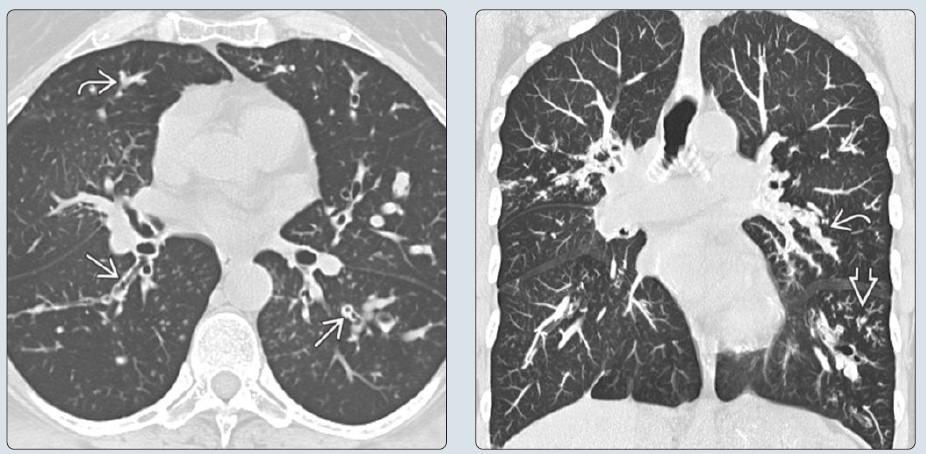
(Left) Axial NECT of the same patient shows extensive bronchiectasis, bronchial wall thickening ſt, and branching opacities that represent mucoid impactions st. (Right) Coronal NECT MIP image of the same patient shows bronchiectasis, mucoid impactions st, and tree-in-bud opacities from bronchiolar mucoid impactions. Cylindrical bronchiectasis is more common in asthma, and cystic or varicoid bronchiectasis is more common in allergic bronchopulmonary aspergillosis. (Courtesy S. Rossi, MD.)
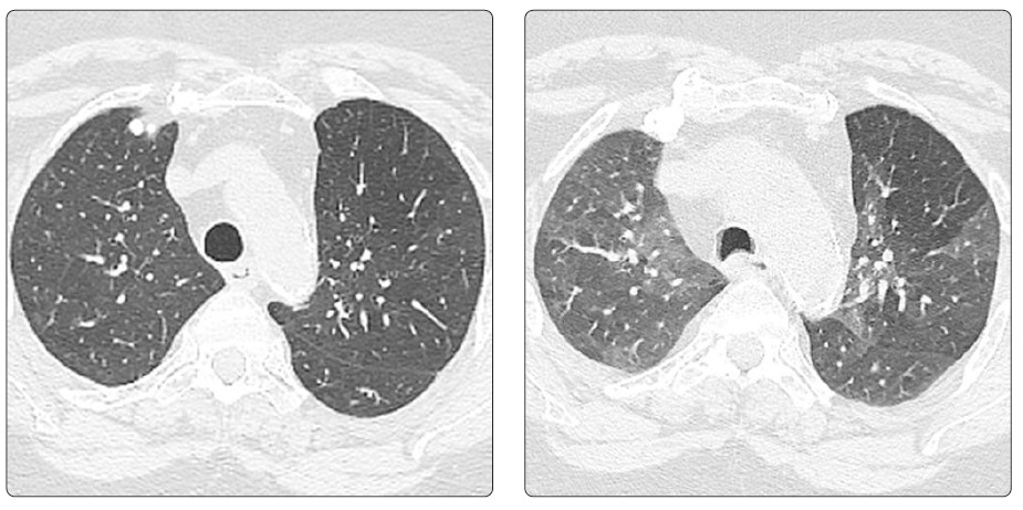
(Left) Axial inspiratory HRCT of a patient with asthma shows a very subtle pattern of bilateral mosaic attenuation. (Right) Axial expiratory HRCT of the same patient shows scattered bilateral subsegmental air-trapping, consistent with small airways disease. This finding often correlates with severity of asthma and is associated with a history of asthma-related hospitalization, intensive care unit admissions, &/or mechanical ventilation.
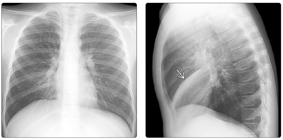
(Left) PA chest radiograph of a young patient with asthma shows mild elevation of the right hemidiaphragm and obscuration of the right cardiac border secondary to atelectasis of the middle lobe. (Right) Lateral chest radiograph of the same patient shows a band-like opacity ſt caudal to the inferiorly displaced horizontal fissure that confirms middle lobe atelectasis. Atelectasis is one of the most common abnormalities found on chest radiographs of patients with asthma.
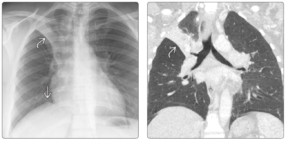
(Left) PA chest radiograph of a patient with asthma shows a right upper lobe opacity with elevation of the minor fissure st and a juxtaphrenic peak ſt, consistent with right upper lobe atelectasis. (Right) Coronal CECT of the same patient shows sublobar right upper lobe atelectasis st. Atelectasis in patients with asthma is typically associated with mucous plugs and does not necessarily imply acute illness, infection, or worsening asthma.
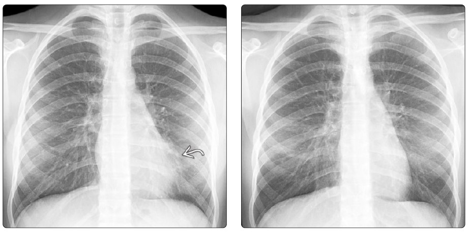
(Left) PA chest radiograph of a patient with asthma who presented with dyspnea, fever, and leukocytosis shows obscuration of the left heart border st, consistent with lingular pneumonia given the history. (Right) PA chest radiograph of the same patient shows post-treatment resolution of the lingular consolidation. Pneumonia is a common complication of asthma and an indication for imaging asthmatic patients. Since asthma is so prevalent, an effort should be made to image affected patients as little as possible.
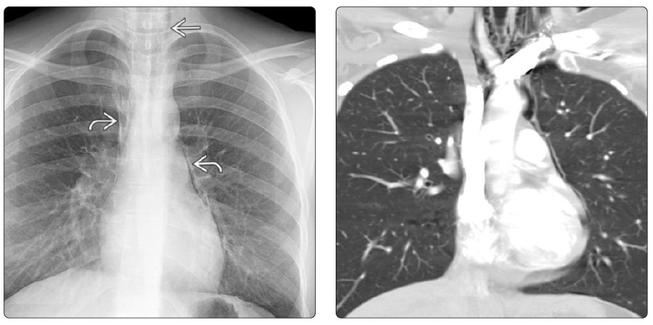
(Left) PA chest radiograph of a patient with asthma who presented with acute dyspnea and chest pain shows extensive pneumomediastinum st and subcutaneous air ſt in the neck. (Right) Coronal CECT of the same patient shows pneumomediastinum and subcutaneous air in the neck. Pneumomediastinum as a complication of asthma is more common in children and more frequent than pneumothorax. Rarely, pneumomediastinum may be associated with air within the spinal canal.
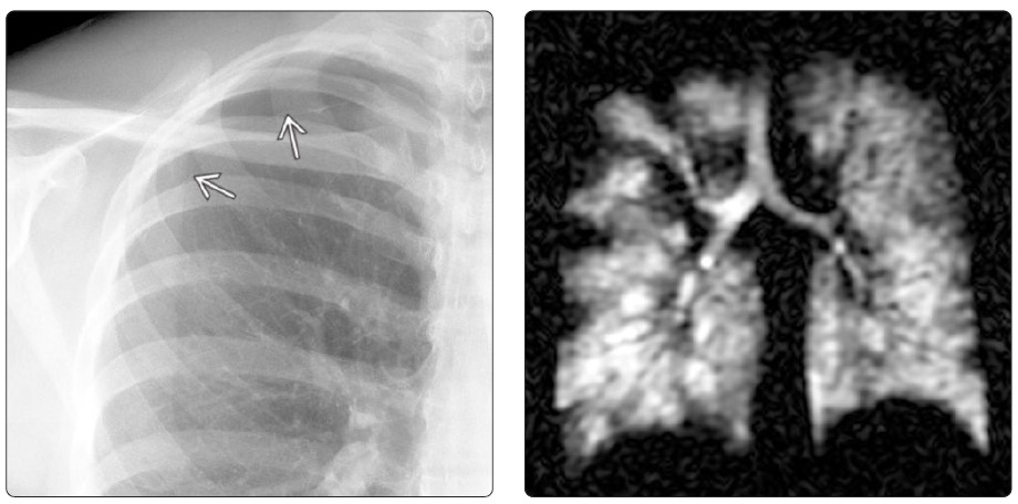
(Left) PA chest radiograph of a patient with asthma shows a pneumothorax manifesting with a visible visceral pleural line ſt at the right lung apex. (Right) Coronal MR with hyperpolarized ¹²⁹Xe of an asthmatic patient shows heterogeneous distribution of ¹²⁹Xe due to extensive ventilation defects. ³He and ¹²⁹Xe have been used successfully in research studies designed to assess ventilatory abnormalities and are promising techniques for future clinical practice. (Courtesy H. P. McAdams, MD.)
Commercial reuse of this material not permitted. Any educational reuse of these images or pdfs must include the following:
Courtesy [insert author names], copyright Elsevier, 2021, reused with permission.