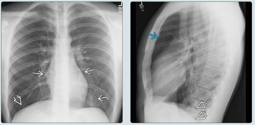
(Left) Frontal radiograph in a 17-year-old during an asthma attack shows hyperexpanded lungs, resulting in a narrow cardiac silhouette ſt & flattening of the diaphragm . Subsegmental opacity in the left lower lobe st was favored to represent atelectasis. (Right) Lateral image in the same patient reveals prominence of the retrosternal clear space & diaphragmatic flattening caused by air trapping. Imaging is often near-normal in asthma patients & is best used to identify complications or alternative diagnoses.
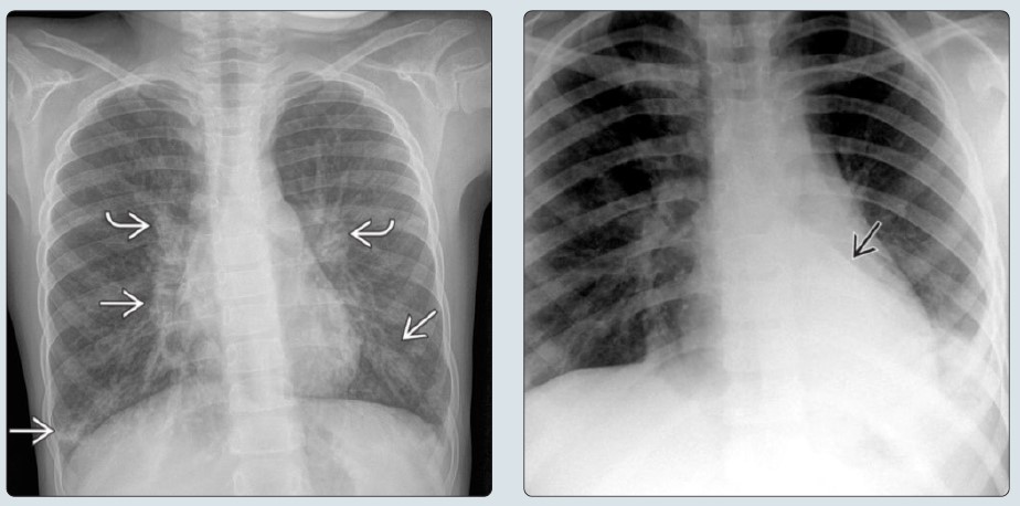
(Left) Frontal radiograph in a 6-year-old with an asthma exacerbation caused by Mycoplasma pneumoniae shows peribronchial airway thickening st & scattered subsegmental opacities in the perihilar & lower lungs ſt. Infection from M. pneumoniae is a common cause of asthma exacerbation. (Right) Frontal radiograph in a child with shortness of breath shows a dense, triangular-shaped opacity from left lower lobe collapse. Lobar collapse & atelectasis are common findings in patients with asthma exacerbation.
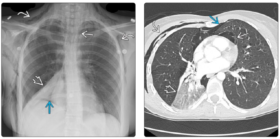
(Left) Frontal radiograph in a 17-year-old with a severe asthma attack precipitated by rhinovirus infection shows extensive subcutaneous emphysema st, pneumomediastinum ſt, right pneumothorax, & right lower lobe collapse. (Right) Axial CECT in the same patient shows extraventilatory gas in the soft tissues st & mediastinum ſt as well as a small left pneumothorax & right lower lobe collapse. Barotrauma is a common complication in asthma patients.
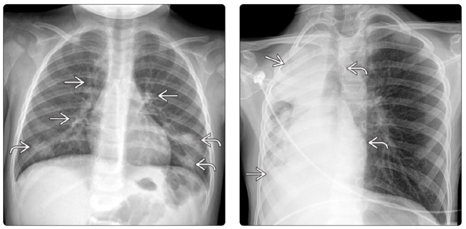
Left) Frontal chest radiograph from a young child with wheezing (who was later diagnosed with asthma) shows peribronchial airway thickening ſt & multifocal atelectasis st bilaterally. (Right) Frontal radiograph from a child with chronic asthma complicated by plastic bronchitis (which is the development of luminal casts, in this case due to inflammation) that caused bronchial occlusion & near total right lung collapse shows widespread opacity ſt & shift of the mediastinum to the right st.
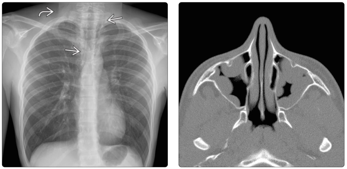
(Left) Frontal chest radiograph in a 14-year-old with a severe asthma exacerbation shows subcutaneous emphysema in the neck st & streaky lucencies in the mediastinum ſt from pneumomediastinum. (Right) Axial sinus CT in a 14-year-old with asthma & allergic rhinitis reveals extensive sinonasal polyposis. The united airway disease hypothesis underscores how sinonasal disease & airway hyperreactivity seen in asthma are manifestations of the same inflammatory process.
Commercial reuse of this material not permitted. Any educational reuse of these images or pdfs must include the following:
Courtesy [insert author names], copyright Elsevier, 2021, reused with permission.