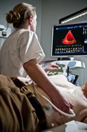
Echocardiogram
An echocardiogram is a test that uses sound waves to make images of your heart. This way of making images is often called ultrasound.
The images from this test can help find out many things about your heart, including:
The size and shape of your heart.
The strength of your heart muscle and how well it's working.
The size, thickness, and movement of your heart's walls.
How your heart valves are working.
- Problems such as:
A tumor or a growth from an infection around the heart valves.
Areas of heart muscle that aren't working well because of poor blood flow or injury from a heart attack.
An aneurysm. This is a weak or damaged part of an artery wall. An artery is a blood vessel.
Tell a health care provider about:
-
Any allergies you have.
-
All medicines you're taking, including vitamins, herbs, eye drops, creams, and over-the-counter medicines.
-
Any bleeding problems you have.
-
Any surgeries you've had.
-
Any medical problems you have.
-
Whether you're pregnant or may be pregnant.
What are the risks?
Your health care provider will talk with you about risks. These may include an allergic reaction to IV dye that may be used during the test.
What happens before the test?
You don't need to do anything to get ready for this test. You may eat and drink normally.
What happens during the test?

-
You'll take off your clothes from the waist up and put on a hospital gown.
-
Sticky patches called electrodes may be placed on your chest. These will be connected to a machine that monitors your heart rate and rhythm.
-
You'll lie down on a table for the exam. A wand covered in gel will be moved over your chest.
- Sound waves from the wand will go to your heart and bounce back—or "echo" back. The sound waves will go to a computer that uses them to make images of your heart. The images can be viewed on a monitor.
-
You may be asked to change positions or hold your breath for a short time. This makes it easier to get different views or better views of your heart.
-
In some cases, you may be given a dye through an IV. The IV is put into one of your veins. This dye can make the areas of your heart easier to see.
The procedure may vary among providers and hospitals.
What can I expect after the test?
-
You may return to your normal diet, activities, and medicines unless your provider tells you not to.
-
If an IV was placed for the test, it will be removed.
-
It's up to you to get the results of your test. Ask your provider, or the department that's doing the test, when your results will be ready.
This information is not intended to replace advice given to you by your health care provider. Make sure you discuss any questions you have with your health care provider.
