Tetralogy of Fallot, Pediatric
Tetralogy of Fallot is a combination of four heart defects that are present at birth (congenital). The condition develops in an unborn baby early in pregnancy, while the heart is developing. Together, these defects cause problems in blood flow, which makes it difficult for the body to get enough oxygen-rich blood.
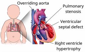
The four heart defects that make up tetralogy of Fallot are:
Ventricular septal defect. This is a hole in the wall between the two lower chambers of the heart (ventricles). The hole allows blood with oxygen to mix with blood without oxygen.
Overriding aorta. Normally, the aorta is attached to the left ventricle. In an overriding aorta, it is located between the ventricles and over the ventricular septal defect. An overriding aorta allows the oxygen-poor blood from the right ventricle to travel to the rest of the body.
Pulmonary stenosis. This is a condition in which the valve between the right ventricle and the artery that leads to the lungs (pulmonary artery) is too narrow. Pulmonary stenosis results in less blood moving to the lungs than is normal.
Right ventricular hypertrophy. This is an enlargement and thickening of the right ventricle. It occurs because the heart has to work harder than normal to move blood through the pulmonary artery.
Tetralogy of Fallot is usually discovered within the first year of life when a health care provider hears an abnormal sound (heart murmur) when listening to the heart beat.
What are the causes?
The cause of this condition is not known. In some cases, it may be caused by genes that are passed from parent to child (inherited).
What increases the risk?
This condition is more likely to develop in children who have DiGeorge syndrome or Down syndrome. It is also more likely to develop during pregnancy if the child's mother:
What are the signs or symptoms?
Symptoms of this condition include:
A bluish tint to the skin, fingertips, or lips (cyanosis).
Difficulty breathing.
Being tired or limp.
Not responding when touched or being talked to.
Fussiness.
A seizure.
When these symptoms occur suddenly, it is called a tetralogy spell. Tetralogy spells occur when there is a sudden drop in blood oxygen levels.
How is this diagnosed?
This condition is diagnosed with a test that uses sound waves to produce images of the heart (
echocardiogram). Additional tests may be done to confirm the diagnosis. These may include:
A physical exam.
Chest X-ray.
Electrocardiogram (ECG) to check for irregular heartbeats (arrhythmias).
Echocardiogram to check the child's heart muscle function.
Blood tests.
Pulse oximetry to check the amount of oxygen in the blood.
An exercise stress test. During this test, your child exercises or is given medicine to make the heart work harder and beat faster while heart tests are done.
How is this treated?
Your child will see a heart specialist (
cardiologist) to manage this condition. The condition is treated with:
- Open-heart surgery. This is done within the first year after birth. The surgery is done to:
Close the ventricular septal defect.
Widen the pulmonary artery.
Correct the problems that are caused by the other defects.
- Shunting. This temporary surgery is performed on children who cannot have open-heart surgery for any reason, such as those who are very small for their age. The purpose of the surgery is to:
Place a device called a shunt between the pulmonary artery and the aorta.
Create a pathway between the pulmonary artery and the aorta, allowing more blood to travel to the lungs.
This information is not intended to replace advice given to you by your health care provider. Make sure you discuss any questions you have with your health care provider.