
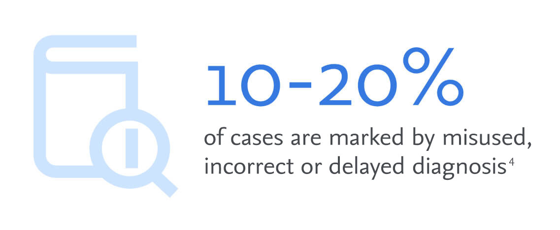
To help alleviate the pressure, to quickly and accurately diagnose each radiology case, as imaging volumes increase, Elsevier have developed Pinpoint cases.
A resource that shares practical expertise and insights using reference cases from renowned radiology experts in their field. Our aim is to help radiologists at all levels, tackle the workloads they encounter each day in a more efficient way and find answers faster, to many diagnostic dilemmas.
Cases from many subspecialities including Pediatrics, Genitourinary, Gastrointestinal and Breast imaging
Expert led examples from renowned radiology specialists
Explore the techniques that experts use to increase speed to diagnosis
Reduce unnecessary tests with a more robust diagnostic process
Learn how to use diagnostic tools experts favor
Pediatrics
Learn how to recognize peadiatric soft tissue masses – Infantile Hemangioma
Genitourinary
Learn how to recognize the different patterns for Prostate Gland Anatomy on MRI
Genitourinary
Learn how to identify the different patterns for assessing prostate anomalies using MRI sequences
Breast imaging
Learn how to recognise the different patterns for assessing breast microcalcifications.
Gastrointestinal
Learn how to identify Cystic Lesions of the Pancreas
GASTROINTESTINAL
Learn imaging techniques for CT & MR Enterography
Diverticulitis
Learn imaging techniques for Diverticulitis
Contact us today
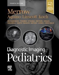
Merrow | Aquino | Linscott | Koch

Fananafazir | Foster
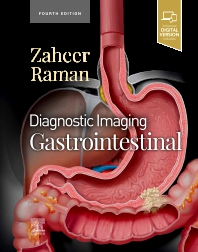
Zaheer | Raman
Dr. Matthew Morgan specializes in diagnostic radiology in Salt Lake City, UT and has over 19 years of experience in the field of medicine. He graduated from University of Utah School of Medicine with his medical degree in 2003. He is affiliated with numerous hospitals in Utah and more, including University of Utah Hospitals and Clinics. Dr. Matthew Morgan is licensed to practice by the state board in Utah (7280832-1205).
Dr. Matthew Morgan is also the author of numerous peer-reviewed articles and textbook chapters and has earned multiple national research awards. Dr. Matthew Morgan specializes in the interface between medicine and technology and work to create systems that help doctors make better decisions at the point of care.
Carl Merrow attended medical school at the University of Alabama School of Medicine and subsequently completed a radiology residency at the University of Alabama at Birmingham. Dr. Merrow then spent two years in fellowship at Cincinnati Children’s Hospital Medical Center. The first year was devoted to general pediatric radiology while the second year was divided between pediatric musculoskeletal imaging and pediatric neuroradiology.
After working at The Children’s Hospital of Alabama for two years, Dr. Merrow returned to Cincinnati Children’s Hospital as a Staff Radiologist and Assistant Professor of Radiology at the University of Cincinnati College of Medicine. He specializes in pediatric musculoskeletal imaging and fetal imaging. Particular interests include vascular malformations, musculoskeletal neoplasms,
Dr. Bryan Foster is an Assistant Professor in the Body Imaging Section at Oregon Health & Science University. He grew up in the United States Northeast and attended medical school at Boston University.
Dr. Foster stayed on at Boston University/Boston Medical Center for his radiology residency where he was Chief Resident. He then completed an abdominal imaging fellowship at the University of Utah.
Dr. Atif Zaheer is an associate professor of radiology and radiological science at Johns Hopkins University School of Medicine, as well as an associate program director to the diagnostic radiology residency. Dr. Zaheer also serves as the director of the Cross-Sectional Body Imaging Fellowship program. Dr. Zaheer is board certified in diagnostic radiology and practices at Johns Hopkins Hospital in Baltimore, MD. and is also affiliated with Sibley Memorial Hospital and Sibley’s Prostate Cancer Multidisciplinary Clinic in Washington, D.C.
Dr. Zaheer earned his medical degree from the Aga Khan University. He completed a residency in diagnostic radiology at Beth Israel Deaconess Medical Center/Harvard Medical School, followed by a fellowship in abdominal imaging and interventional radiology at Brigham and Women’s Hospital/Harvard Medical School.Dr. Zaheer is clinically active in cross-sectional imaging of the body using CT, MRI and Ultrasound and is also involved in multidisciplinary conferences for pancreas, liver and rectal cancer imaging. His research interests include imaging of tumors and inflammatory disorders of the pancreas and is considered a leader in pancreatic imaging.
Ben Wildman-Tobriner is an associate professor of radiology at Duke University in Durham, North Carolina. He is director of the abdominal imaging fellowship program at Duke, and has research interests in liver imaging and thyroid ultrasound. He has lectured around the United States on a variety of radiology topics including gastrointestinal pathology, contrast-enhanced ultrasound, and cognition in radiology.
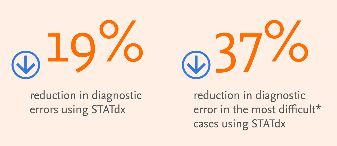
STATdx, as featured in our expert cases, is a valuable diagnostic workflow tool providing decision support in helping to reduce diagnostic errors and is especially valuable when diagnosing rare or complex imaging cases.
Cookies are used by this site. To decline or learn more, visit our cookie notice.
Copyright © 2025 Elsevier, its licensors, and contributors. All rights are reserved, including those for text and data mining, AI training, and similar technologies.